


Pallas Bugseed Possibly Extirpated in Manitoba

Pallas Bugseed Possibly Extirpated in Manitoba

For the last two years I have been searching for four species of rare Bugseed (Corispermum spp.) plants. Historically these plants were found in sand dunes and along the beaches of Lakes Winnipeg and Manitoba. Unfortunately there were very few recently collected specimens; most had been collected over 40 years ago. Attempts to determine the rarity status of these plants were hampered due to this lack of information.

During my field work I relocated fewer than half of the historical populations of Bugseeds indicating a substantial loss of habitat. In some cases, dune stabilization appears to have resulted in the loss of habitat for these species, which grow in bare sand. Flooding along the Red River and on Lakes Manitoba and Winnipeg in 2010 and 2011 may have also caused the loss of habitat as several historical populations had been previously found on the beaches, dunes and sandbars along the south shore of these lakes. Balancing out the losses a bit was the discovery of seven new localities in the province including a very large population of Hairy Bugseed (C. villosum) in the sand dunes in Grand Beach Provincial Park.
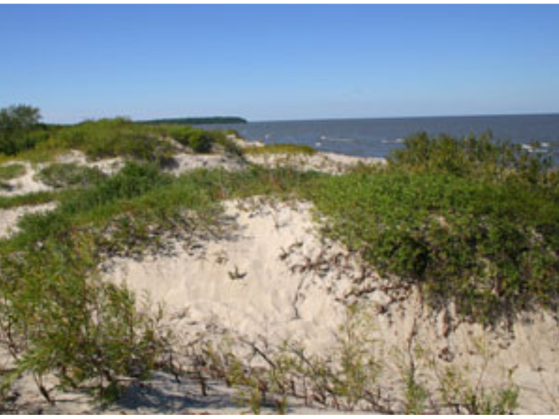
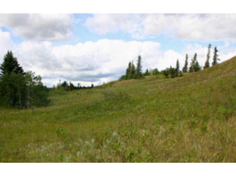
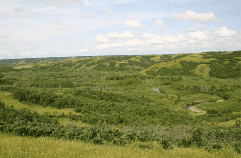
A series of sand dunes with grass growing at the tops. A body of water is partially visible in the distance beyond the dunes.

A new population of Hairy Bugseed was discovered at Grand Beach!
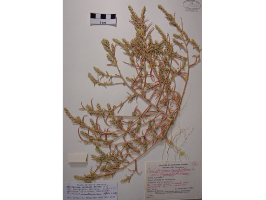
A dried herbarium specimen of Pallas Bugseed with specimen details in the bottom right of the base paper.

Pallas Bugseed has not been seen in Manitoba in over 60 years!

Unfortunately, I could not locate any Pallas Bugseed (C. pallasii) at the three sites where it was historically found although I did find some American Bugseed (C. americanum) at two of the sites. Pallas Bugseed is still present in other Canadian provinces so it is not extirpated in the country. It is possible that this species spread into Manitoba from other provinces where it is native along railways and then subsequently disappeared as more hardy weeds took over the habitat. As the seeds of these species are quite long lived and tolerant of burial, Pallas Bugseed may still be here in the province, hiding in the soil and waiting for the right conditions to germinate.

Dr. Diana Bizecki Robson

Dr. Diana Bizecki Robson

Curator of Botany

Dr. Bizecki Robson obtained a Master’s Degree in Plant Ecology at the University of Saskatchewan studying rare plants of the mixed grass prairies. After working as an environmental consultant and sessional lecturer…
Meet Dr. Bizecki Robson

Holding it Together

The Canadian Conservation Institute (CCI) is part of the federal Department of Canadian Heritage, created to promote the preservation of Canada’s cultural heritage. Every two to three years, CCI develops and hosts in Ottawa a symposium on a conservation topic.

For a week in October conservator Lisa May attended Symposium 2011 – Adhesives and Consolidants for Conservation: Research and Applications.

Internationally attended, the symposium covered the newest research, techniques and products for a wide range of adhesives and consolidants used in conservation. Papers and posters addressed use of these products on virtually every type of material that conservators encounter.

A dedicated poster session and generous breaks allowed for opportunity to speak to the presenters as well as mingle with the over 200 participants. In addition, one afternoon was dedicated to tours of Ottawa institutions.

The last day of the symposium was at CCI where the paper authors, as well as other conservators and scientists working at CCI, demonstrated their new research, techniques, and applications. There were over 30 participants presenting 25 minute demonstrations, with each offered four to five times during the day to accommodate all those in attendance. -Lisa May

The opportunity to attend this symposium has allowed for Lisa to return with new contacts and up-to-date information we will use in conservation treatments. In particular, there are some Natural History specimens awaiting our attention, about which she gleaned pertinent suggestions on materials and techniques. Below are three photos Lisa took during the demonstration day.

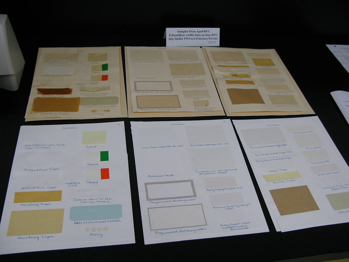
Six sheets of paper, the top three darkens to cream, and the bottom three white. All have various tape samples on them.

Original and oven aged self-adhesive tapes and labels samples.

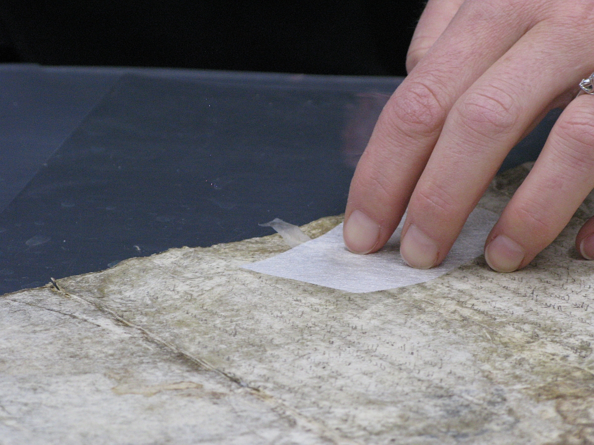
Close up on somebody's hand as they repair a torn parchment.

Repairing a parchment tear with gelatine and goldbeater’s skin.

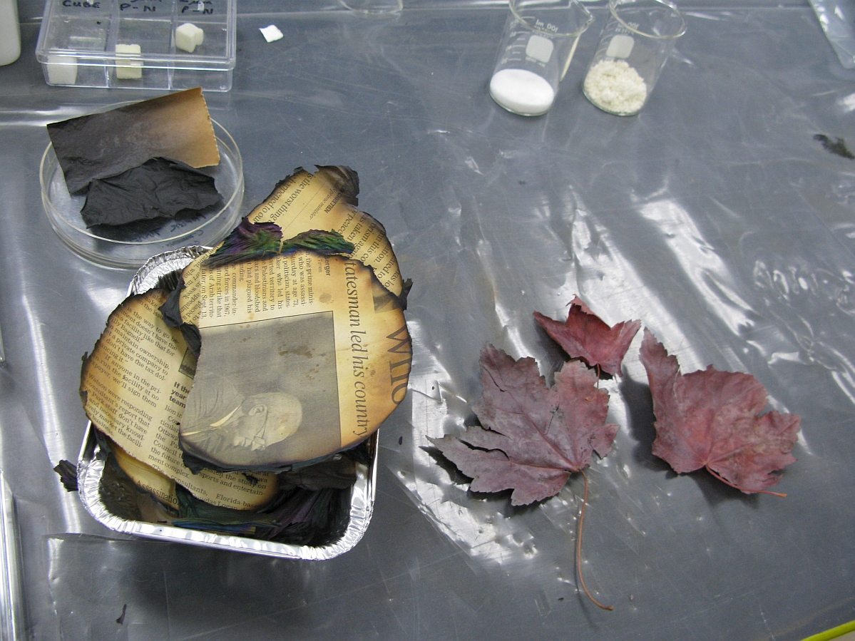
A small container with pieces of newspaper next to three red leaves. Both types of objects have charred edges.

Parylene coated newspapers and leaves.

The Hong Kong Veterans, 1941-1945

Part II

During the Battle of Hong Kong, 290 of the 1,975 Canadians defending the island were killed in battle. After the Canadians were captured by the Japanese on Christmas Day, 1941, Canadian soldiers were taken into a brutal period of captivity, first in Hong Kong and then in Japan. Deprived of food and sanitary conditions, 267 more Canadians died as Prisoners of War.

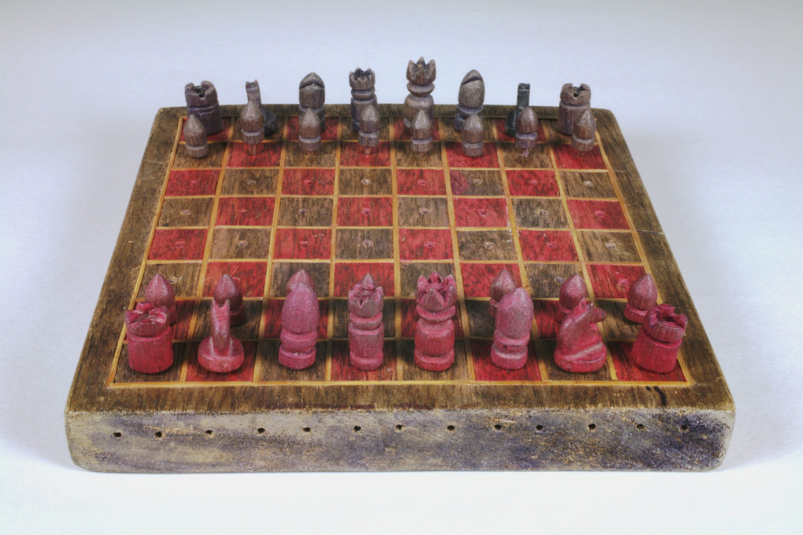
A rough, handmade chess set with red and platinum coloured pieces lined up in starting positions.

In Hong Kong the Winnipeg Grenadiers suffered through long days of hunger and boredom. Woodworking contests were set up to keep minds and hands busy. A very recent donation to the Manitoba Museum includes one of these wooden artifacts: a hand-carved chess set inlaid with bamboo. This belonged to Lieutenant Richard Maze, who signed up for the Saskatchewan regiment with Corrigan (see Part I here): they were both later moved to the Winnipeg Grenadiers. The complete set features tiny chess pieces (about 2 cm tall) that include thin pegs to secure them to the board. Lieutenant Maze received the set from a fellow prisoner who constructed it from wood scraps found around the Kawloon POW Camp, Hong Kong. This little chess set is an example of how creative activity and friendship helped the prisoners withstand deprivation in such difficult conditions. Thanks to Rose-Ann Lewis and Ann Maze for the donation of Lieutenant Maze’s Hong Kong Veterans items to the Manitoba Museum.


Image: Chess set made by Winnipeg Grenadier POW, Hong Kong, ca. 1942-1944. H9-37-547-a-ag. Unless otherwise noted, The Manitoba Museum holds copyright to the material on this site.

The Canadians were later moved to a POW camp in Japan, where many worked in mines and they were limited to less than 800 calories of food a day.

The Japanese government recently offered a full apology for the treatment of Canadians in these POW camps. (Read a CBC article covering the apology, here).

Reactions among Canadians are mixed, with some accepting the apology while others say it’s too little, too late. What do you think?
Dr. Roland Sawatzky

Dr. Roland Sawatzky

Curator of History

Roland Sawatzky joined The Manitoba Museum in 2011. He received his B.A. in Anthropology from the University of Winnipeg, M.A. in Anthropology from the University of South Carolina, and Ph.D. in Archaeology…
Meet Dr. Roland Sawatzky

Musing About Macoun

Today I got to study a plant specimen that had been collected 100 years before I was born in 1872 by one of the most famous Canadian botanists, Dr. John Macoun. Dr. Macoun accompanied the engineer Sanford Fleming to look for a railroad route through the Canadian west and determine the area’s agricultural potential. Macoun collected thousands of plant and animal specimens on his journeys, which involved travelling either by horse or canoe. He collected over 100,000 plants in his lifetime including over 1,000 that were new to science. In total 48 new species were named after him, including Macoun’s buttercup (Ranunuculus macounii).

Touching Macoun’s handwritten label made me wonder what western Canada looked like back then. Just acres and acres of grasses and fragrant wildflowers, and thousands of birds, antelope and deer I imagine. Or maybe it just felt big and empty and utterly overwhelming.

Looking down a railway track with grassy banks on either side.

Macoun and Fleming were trying to determine the route for the western railroad.

Rolling grassy banks with occasional trees at the edges.

Macoun travelled along the Carlton trail through the Brandon Sand Hills.

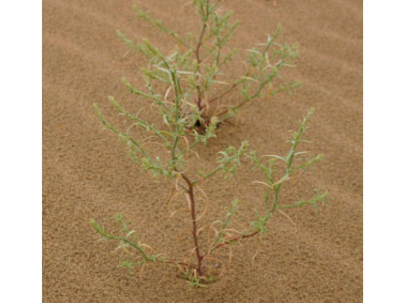
A stringy plant with small green leaves growing in a sand dune.

I can’t imagine the difficulties he encountered trying to do his field work.  Lugging everything you would need for months on pack animals or wagons must have been difficult to manage.  Being a scientist, he also had to protect his plant press full of specimens from insects and moisture, water being a botanists’ greatest enemy.  Many a brave explorer had his precious specimens go completely moldy.  The botanist David Douglas ended up eating part of his plant collection so he wouldn’t starve to death!  The field work that I do is posh in comparison; I drive on paved or gravel roads and usually stay in hotels or cabins!  Furthermore I don’t have a whole chapter at the end of my reports recounting the loss of life that occurred during the field work.  Sanford Fleming’s “Report of progress on the explorations and surveys up to January 1874” contains such a chapter recounting the loss of 19 men during his survey: seven died in a forest fire, four drowned while canoeing the Ottawa River and eight died in a steamer wreck on a shoal!


Image: Dr. Macoun collected a specimen of Hairy Bugseed in 1872 that I have been studying.

I also mused about the end result of his journeys. Macoun’s report on the fertility of the prairies convinced Canada to send immigrant farmers to this land. How would he have felt knowing that his report paved the way for the almost complete destruction of the grassland that he had wandered on? That plants and animals that had once numbered in the billions would be reduced to mere handfuls less than a century after his arrival? Perhaps it would have made him happy, knowing that the land was settled with European farmers and ranchers. But I suspect that that happiness would be mingled with at least some regret that no one would ever again experience the wild west as he had.

Dr. Diana Bizecki Robson

Dr. Diana Bizecki Robson

Curator of Botany

Dr. Bizecki Robson obtained a Master’s Degree in Plant Ecology at the University of Saskatchewan studying rare plants of the mixed grass prairies. After working as an environmental consultant and sessional lecturer…
Meet Dr. Bizecki Robson

The Sloth’s Tale

By Dr. Graham Young, past Curator of Palaeontology & Geology


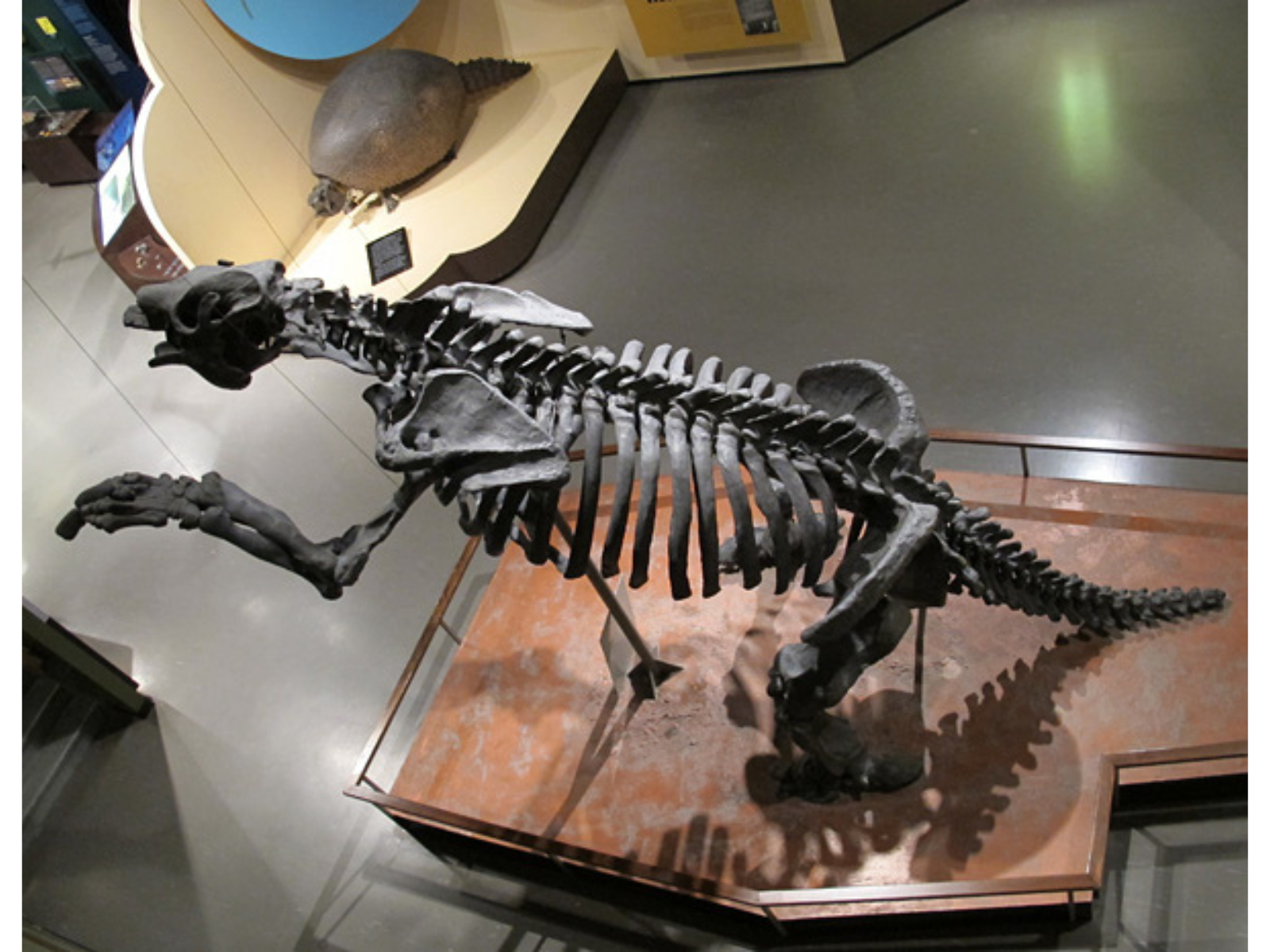
Ever since the Museum’s Earth History Gallery opened in the early 1970s, visitors have been struck by the appearance of a giant skeleton near the end of the gallery. Many children (and perhaps more than a few adults) have thought that it is a dinosaur, but it is in fact a mammal from the much more recent past, a replica of the giant ground sloth Megatherium americanum.

Megatherium was a huge ground-dwelling creature, distantly related to the modern tree sloths. Ground sloths were a very successful group, with fossils known from many parts of South and North America (including western Canada), but sadly they disappeared in the Pleistocene megafaunal extinctions, along with other wondrous creatures such as the woolly mammoth and short-faced bear.

Looking down towards a museum gallery where a large skeleton is posed on its hind legs. In the distance is a skeleton of a creature with a very bulbous rounded shell.

The Megatherium (foreground) and glyptodont (background), viewed from the Earth History mezzanine.

Looking up at a large mounted skeleton posed on its rear legs.

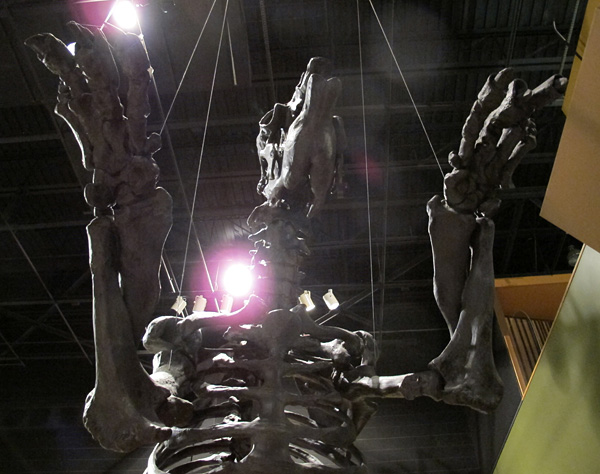
Looking directly up from standing under a large skeleton of a creature with its forearms held up.

There are many different ground sloths known, but Megatherium was the largest, described as “weighing up to eight tons, about as much as an African bull elephant.” It walked on all fours (with a gait similar to that of a giant anteater), but could rise on its hind legs, supported by the huge tail, to browse on the trees that apparently formed the main part of its diet. Our sloth is shown in this sort of pose, though it lacks a tree at present. Although ground sloths lived across both American continents, Megatherium itself is known only from South America.

Our plaster cast of a Megatherium skeleton is, of course, much newer than the Pleistocene. But it is very old in human terms, so remarkably old that it could be considered as an artifact of a long-past scientific age. It is far older than our current Museum building, much older than the first Manitoba Museum that was located in the Winnipeg Auditorium, older than the Manitoba Legislative Building; in fact it is nearly the same age as the Province of Manitoba! The Megatherium and its close colleague the armoured glyptodont have been companions for a century or more, and both arrived at our Museum by a circuitous path.

But let me begin the sloth’s tale at its beginning.

Megatherium is among the best known of ground sloths, with dozens of fossils collected in South America and shipped to Europe from the 18th Century onward. Many specimens apparently came from the banks of the Luján River near Buenos Aires, Argentina, and were in collections and on exhibit in cities such as Paris, Madrid, and London. The first formal scientific description was produced by the great French scientist Georges Cuvier, in 1796. In the 1850s, a young American scientist, Henry Augustus Ward, made a detailed study of Megatherium, visiting collections in Paris and London among many other places. After his return to Rochester, New York, he became a professor at the University of Rochester, but also founded Ward’s Natural Science Establishment, a company that sold scientific specimens, replicas, and materials.

Ward got started on the casting and selling of fossil skeletons very early. His company was among the first to produce casts for sale to the many new natural history museums that were then being developed, and the giant Megatherium was one of his “star attractions.” His catalogue advertised that a full skeleton consisting of 124 different casts could be purchased for $250, “packed not painted” but including a replica tree.

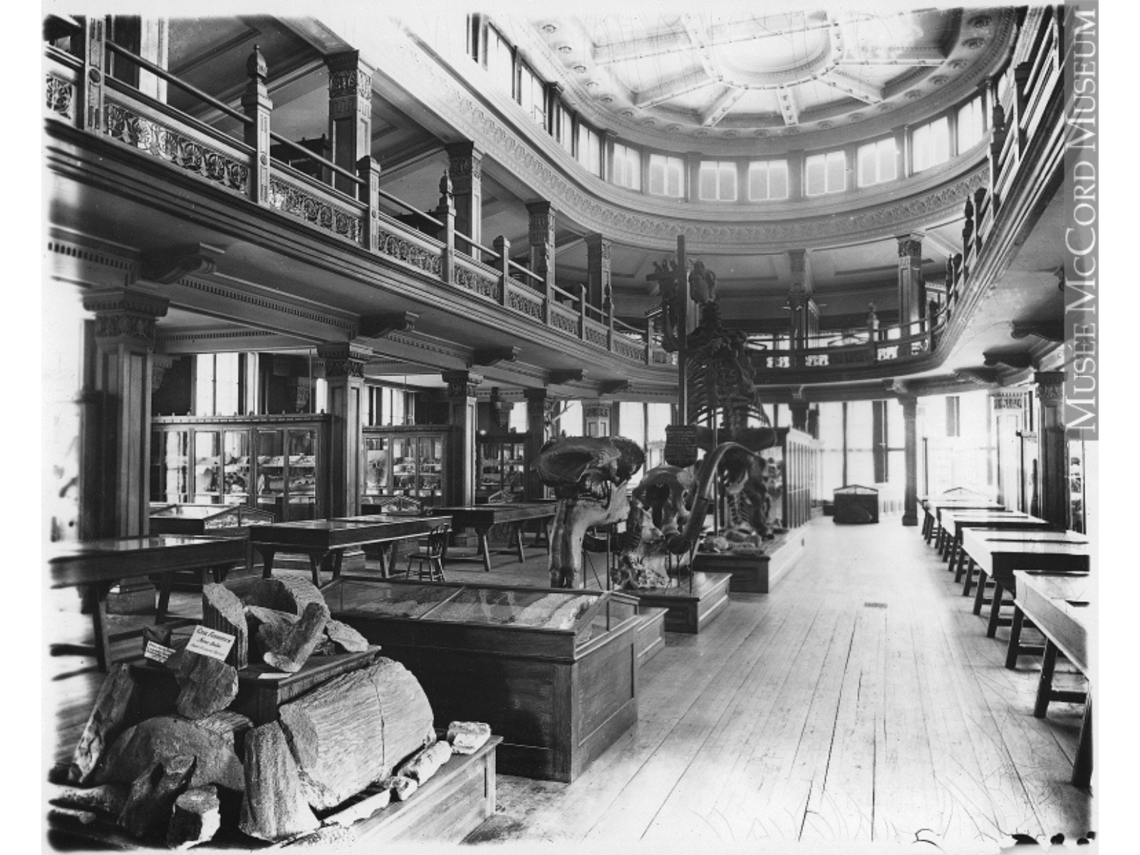
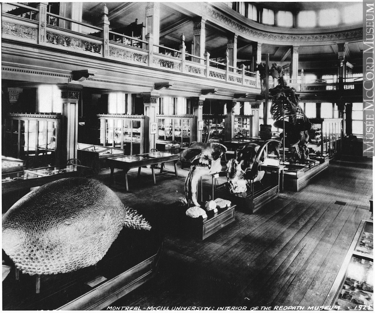
A black and white photo of an old museum. On the ground floor are various display cases as well as a large mounted skeleton posed on their hind legs. A second floor balcony encircles the room's perimeter.

The Redpath Museum c. 1893, with the Megatherium prominently exhibited toward the far end of the gallery. (photo: McCord Museum)

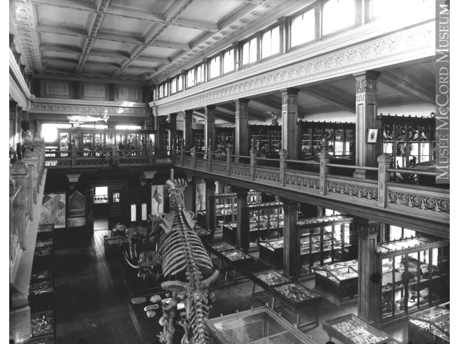
A black and white photo of an old museum taken from a second floor balcony surrounding the room. On the ground floor below are various display cases as well as a large mounted skeleton posed on their hind legs, seen from behind.

View of the Megatherium from above, c. 1893 (photo: McCord Museum)

In Montreal, a museum for McGill University was planned from the 1860s onward, to exhibit collections developed by the world-famous Professor William Dawson. This facility, funded by and named for the industrialist Peter Redpath, was opened in 1882. It was primarily to serve as a resource for the university’s faculty and students, but secondarily for the education of the people of Montreal.

Dawson had long corresponded with Ward concerning the acquisition of particular items, so it is not surprising that a description of the original museum includes:

“Entering the Redpath Museum, the visitor saw at the back of the ground floor a handsome lecture theater with seats for 200 students… To the right of the entrance, a staircase … led to the main floor or “Great Museum Hall.” Henry Ward’s imposing cast of the British Museum’s megatherium (a giant sloth)–set up by his partner Howell and a status symbol for new museums–distinguished this floor, which displayed paleontological, mineralogical, and geological specimens.”
A black and white photo of an old museum. On the ground floor are various display cases as well as a large mounted skeleton posed on their hind legs at the far end of the room. In the foreground is a large four legged creature's skeleton with a bulbous, rounded shell. A second floor balcony encircles the room's perimeter.

The Redpath today is a wonderful old-fashioned natural history museum, but it is also rather pocket-sized in comparison with the huge museums of Europe. The Megatherium occupied a considerable proportion of its limited floor space.  Several years later, it was joined by the armoured glyptodont, also apparently supplied by Ward’s. Thus, that museum’s main hall was dominated by replicas of giant extinct mammals.

But how did these fascinating and historic replicas become migrants once again, moving westward from Montreal to Winnipeg? In the 1960s there must have been a push to add other creatures such as a dinosaur to the Redpath’s exhibits, and the only way to do this was to replace the large pieces that filled its space (that part of the Redpath is now occupied by a tyrannosaurid dinosaur, among other exhibits).


Image: The Redpath in 1925, showing both the glyptodont and the Megatherium. (photo: McCord Museum)

But how did these fascinating and historic replicas become migrants once again, moving westward from Montreal to Winnipeg? In the 1960s there must have been a push to add other creatures such as a dinosaur to the Redpath’s exhibits, and the only way to do this was to replace the large pieces that filled its space (that part of the Redpath is now occupied by a tyrannosaurid dinosaur, among other exhibits).

According to what my predecessor Dr. George Lammers told me, the Redpath was looking for a home for these enormous casts, and this just happened to come at a time when The Manitoba Museum was constructing its new building, with plenty of square footage that needed to be filled. So the skeleton casts were transferred to our Museum, and they were crated and shipped to Manitoba.

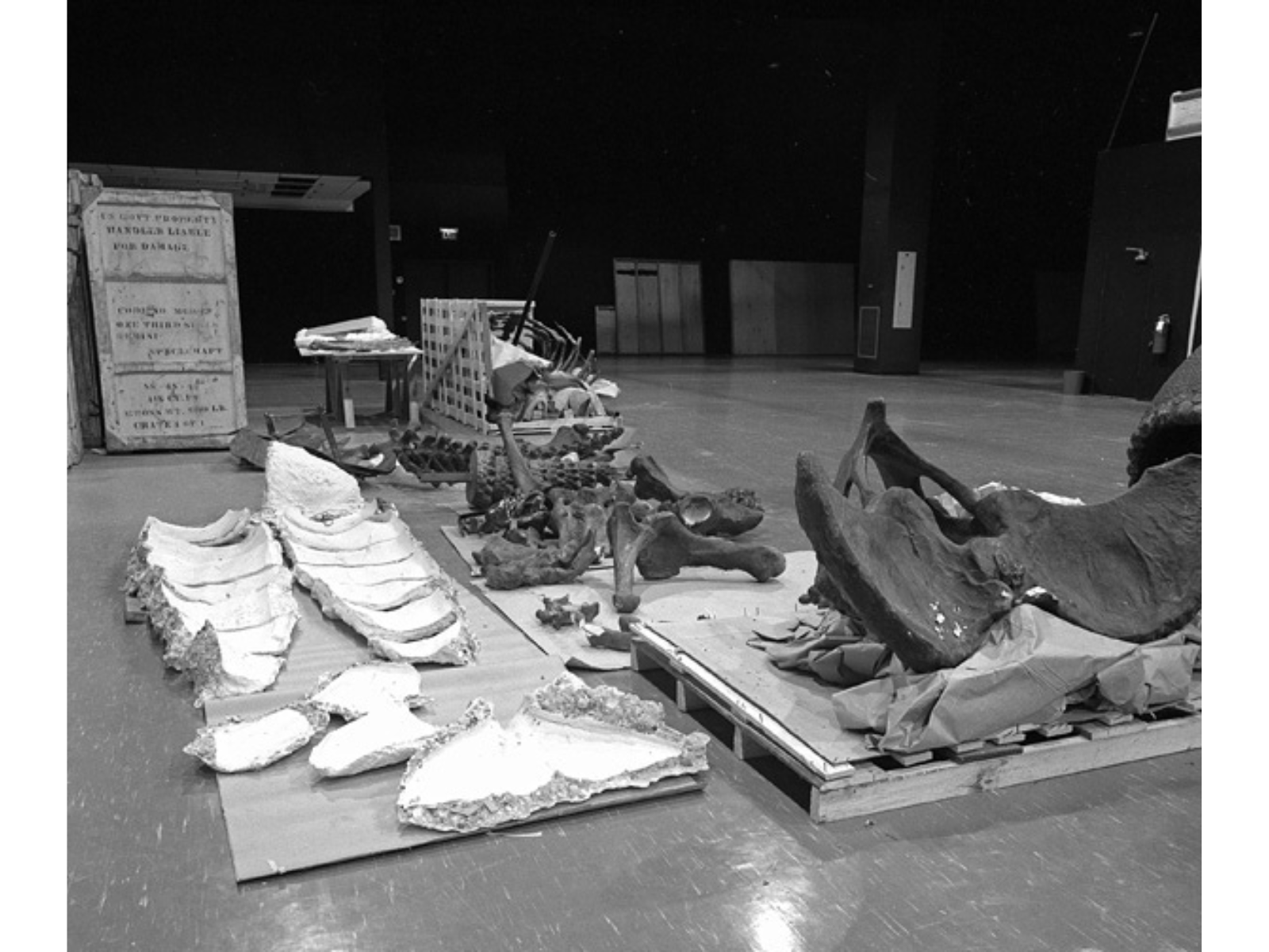
A black and white photo of plaster bones and casts of a large skeleton laid out in pieces in the floor of a mostly empty room.

On arrival at the Museum, the replicas were uncrated and examined. George told me that some repair was required as a result of damage in transit, and apparently the plaster bones were found to be stuffed with Reconstruction Era American newspapers! The Megatherium was re-mounted in a slightly different posture, and sadly lacks the “tree” on which it had appeared to browse for ninety years or so. But at least it had its friend, the glyptodont, nearby, and they seem to have happily settled into their new home, impressing (and sometimes scaring) the millions of children who have visited our Museum in the past forty years.


Image: The disassembled Megatherium, after uncrating but prior to assembly in the as-yet unfinished Earth History Gallery space.

On arrival at the Museum, the replicas were uncrated and examined. George told me that some repair was required as a result of damage in transit, and apparently the plaster bones were found to be stuffed with Reconstruction Era American newspapers! The Megatherium was re-mounted in a slightly different posture, and sadly lacks the “tree” on which it had appeared to browse for ninety years or so. But at least it had its friend, the glyptodont, nearby, and they seem to have happily settled into their new home, impressing (and sometimes scaring) the millions of children who have visited our Museum in the past forty years.

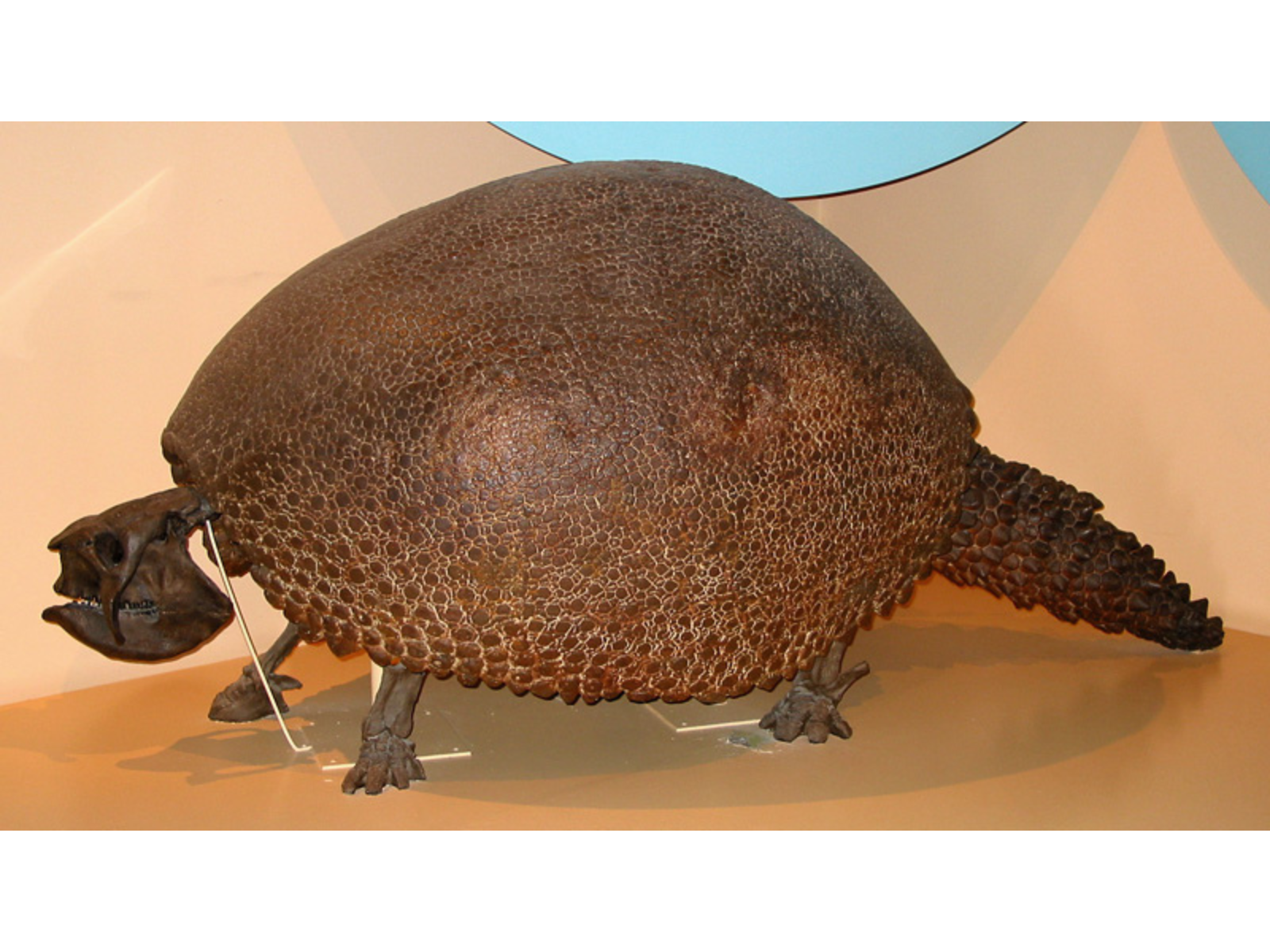
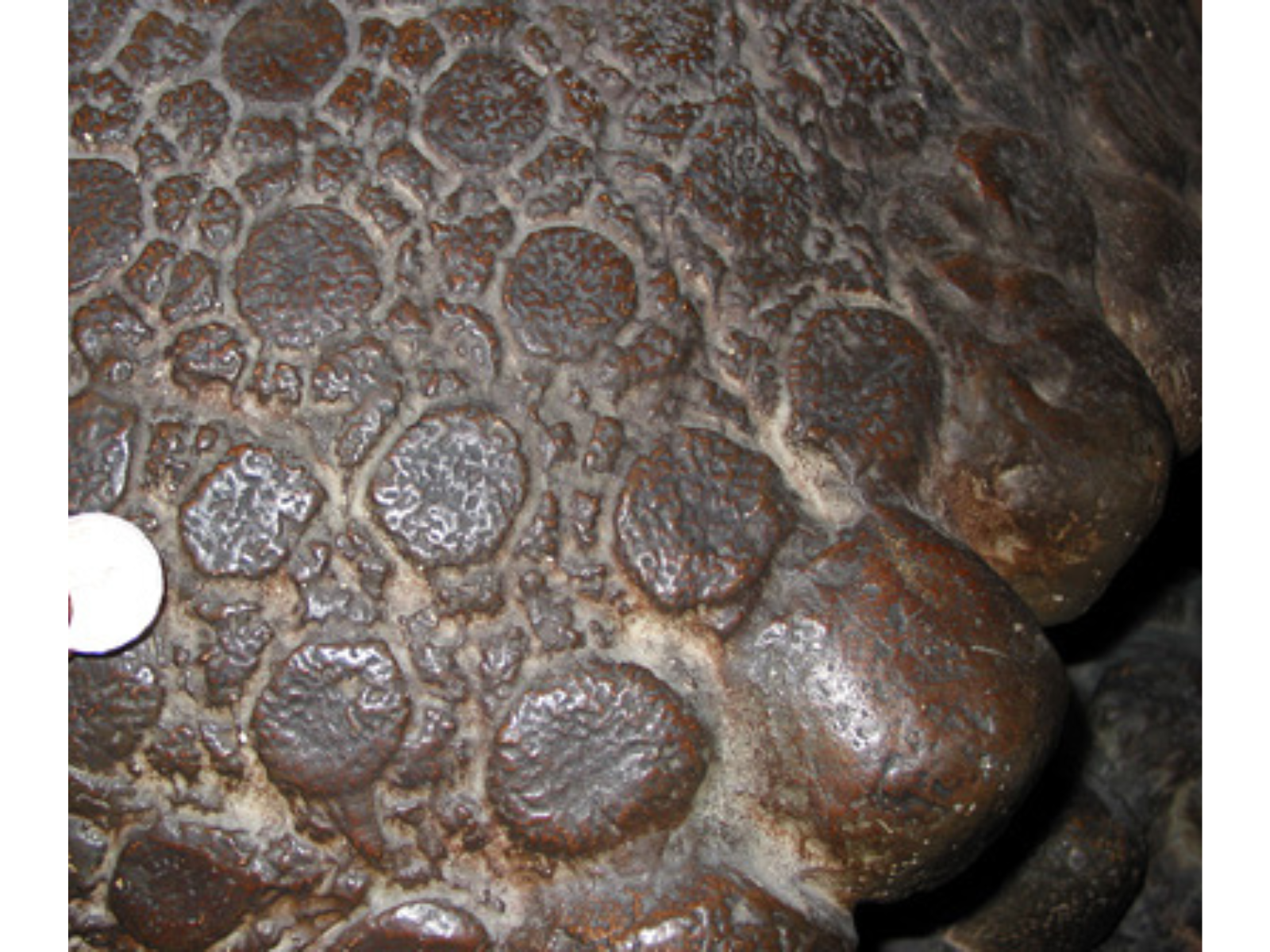
The glyptodont (above) and a detail of the bony plates that made up its protective shell (replica, in our example). Although it looks quite turtle-like, this creature was a mammal related to anteaters, sloths, and armadillos!

A mounted skeleton of a four-legged creature, with a bulbous, rounded shell and a thick tail.

The glyptodont. Although it looks quite turtle-like, this creature was a mammal related to anteaters, sloths, and armadillos!

Close up on a bony plate of shell, covered in irregularly shaped circular marks.

Detail of the bony plates that made up its protective shell (replica, in our example).

The name of our sloth is a bit complicated. The note that George left me calls it Megatherium cuvieri. This is what Ward had called it, and it was probably labelled as such when on exhibit at the Redpath. The species name “cuvieri” was, however, apparently based on a misguided attempt in the 1820s to re-brand Cuvier’s perfectly valid Megatherium americanum. Modern rules of taxonomic usage consider cuvieri to be a nomen illegitimum (“illegitimate name”), so we can  safely call it M. americanum.

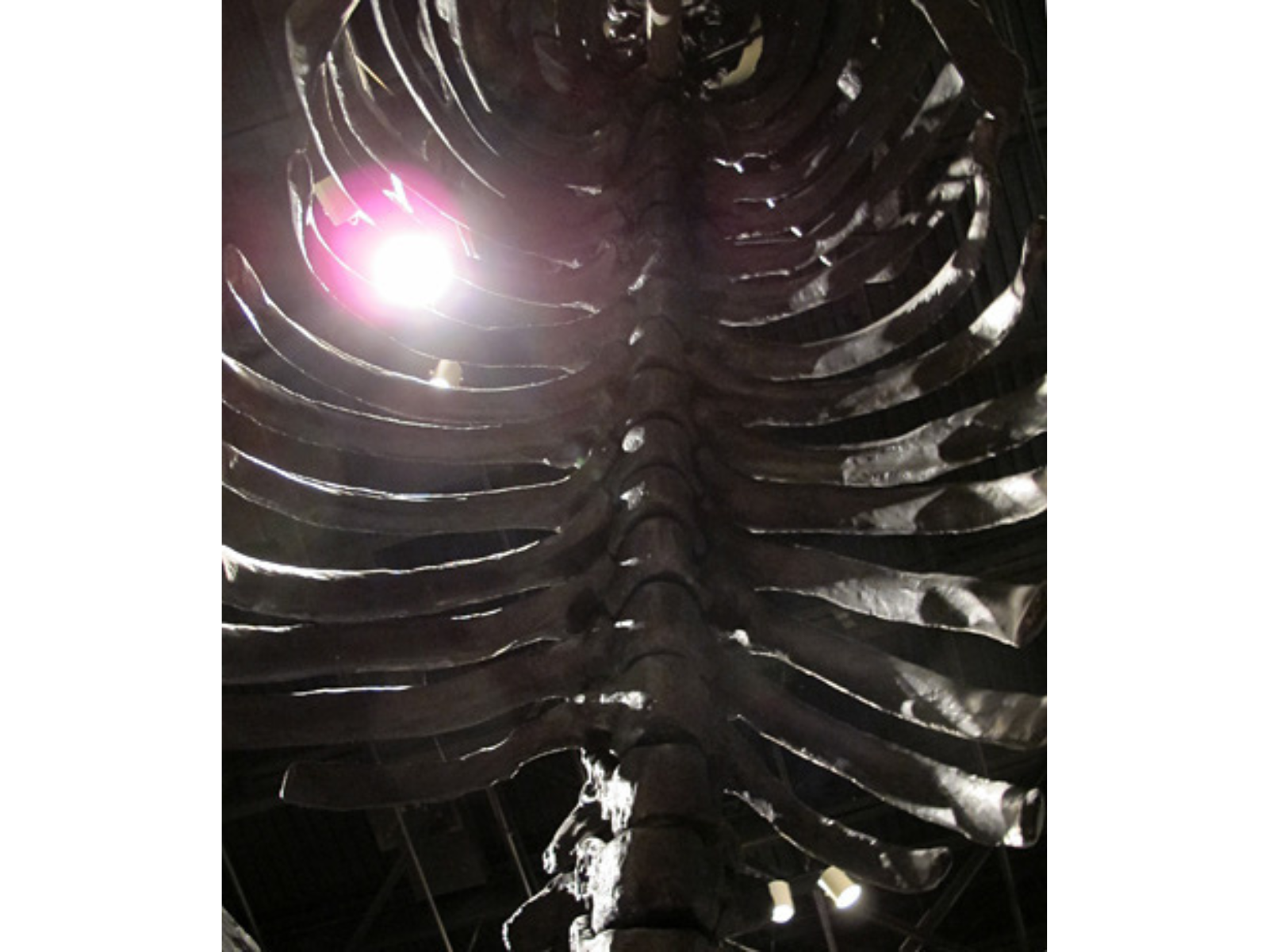
Looking up into the large ribcage of a mounted Megatherium.

The sloth’s massive ribcage.

A large mounted skeleton posed up on its hind legs viewed from behind, as a long tail reaches down from its spine towards the viewer.

The sloth’s tail (of course!).

Given this cast’s critical role in the history of exhibits at two of Canada’s most important natural history museums, and its place in the story of the development of North American paleontology, what is the future of our sloth? Since we have been progressing with a gradual refurbishment of the Earth History Gallery, I would like to soon plan new interpretive materials that explain the tremendous story and significance of this exhibit. But in the somewhat longer term, wouldn’t it be wonderful if it could be re-mounted and given back its “tree,” perhaps serving a new role as the centrepiece for a larger exhibit of extinct ice age animals?

The Winnipeg Grenadiers and the Battle for Hong Kong, 1941

Part I

In the autumn of 1941 World War Two was raging across Europe, but the battles of the Pacific region were yet to come. Although considered of little strategic importance by Winston Churchill, the island of Hong Kong was considered defensible by some Canadian military leaders. On October 20, 1941 the decision was made to send just over 2,000 Canadian soldiers to help defend Hong Kong from possible Japanese aggression. On December 8 the Japanese attacked…

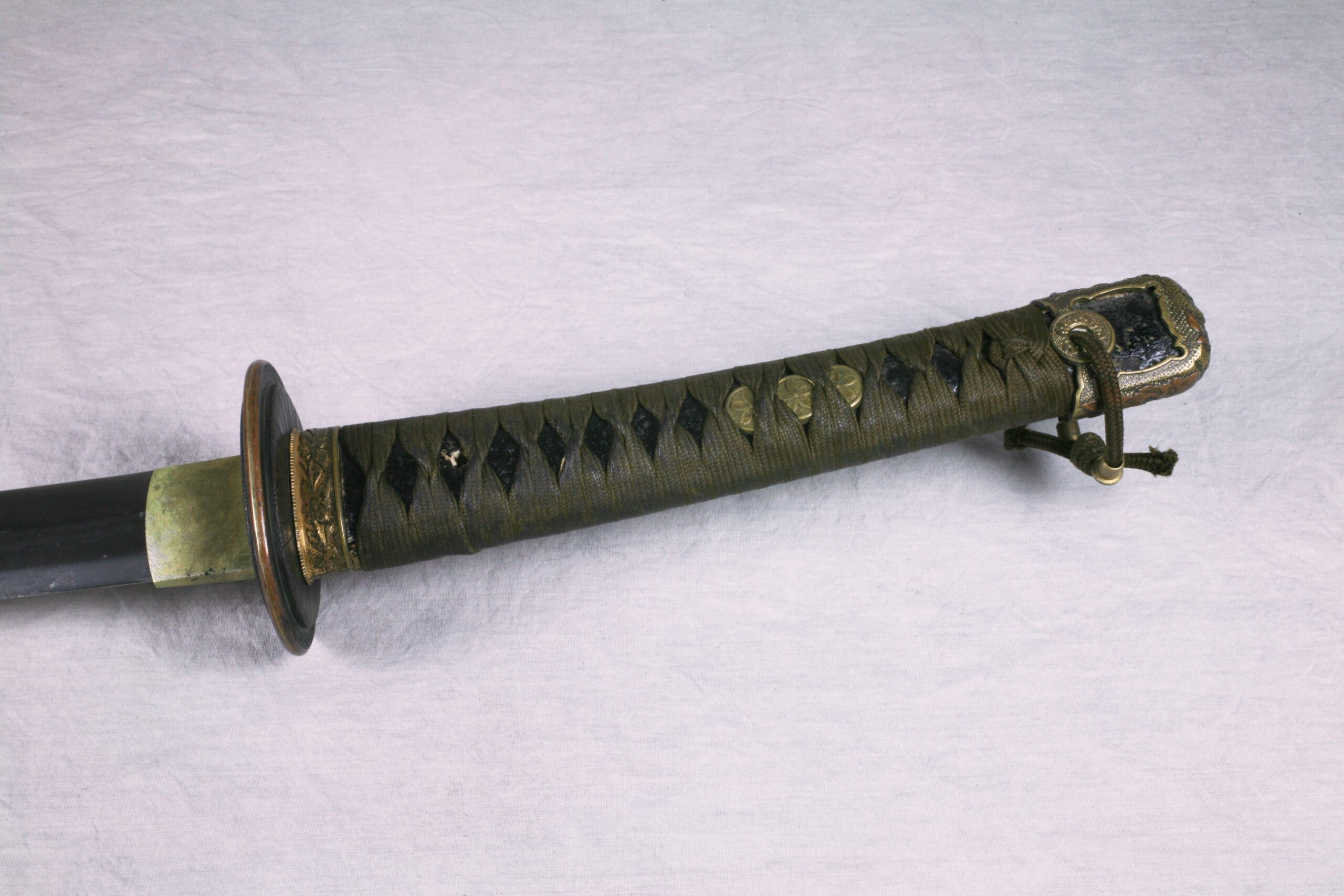
Close up on the hilt and handle of an old Japanese-style military sword.

In defence of the island, the Canadians fought the battle-hardened, well-trained soldiers of the Japanese forces. One artefact of this battle is a Japanese military sword now at The Manitoba Museum. On December 20th, Lieutenant Leonard B. Corrigan was in action with a small fighting patrol of the Winnipeg Grenadiers when they encountered an enemy patrol and engaged in hand-to-hand combat. According to his citation for the award of a Mention-in-Despatches, Corrigan killed two Japanese soldiers and was attacked by a Japanese officer with the sword. He caught the sword with his left hand (suffering a severe injury) and killed the officer with a flare gun. Despite their victory over the enemy patrol, the Canadians were taken prisoner days later and spent the rest of the war in captivity. Their Prisoner of War experience is told at The Manitoba Museum in the Parklands/Mixed Woods Gallery.


Image: Japanese Military Sword (detail of handle) H9-36-184. Unless otherwise noted, the Manitoba Museum holds copyright to the material on this site.
Part Two of this blog will showcase an artefact from their POW experience, when food was in short supply and the days were long.

For more information on the Battle for Hong Kong, visit http://www.hkvca.ca, or for more on the experience of Canadians in the Pacific during the Second World War, visit the exhibit (which will feature some of our artifacts) in Calgary – http://themilitarymuseums.ca/whats-new

Dr. Roland Sawatzky

Dr. Roland Sawatzky

Curator of History

Roland Sawatzky joined The Manitoba Museum in 2011. He received his B.A. in Anthropology from the University of Winnipeg, M.A. in Anthropology from the University of South Carolina, and Ph.D. in Archaeology…
Meet Dr. Roland Sawatzky

New Safety Lines Will Allow Staff to Inspect Nonsuch Rigging

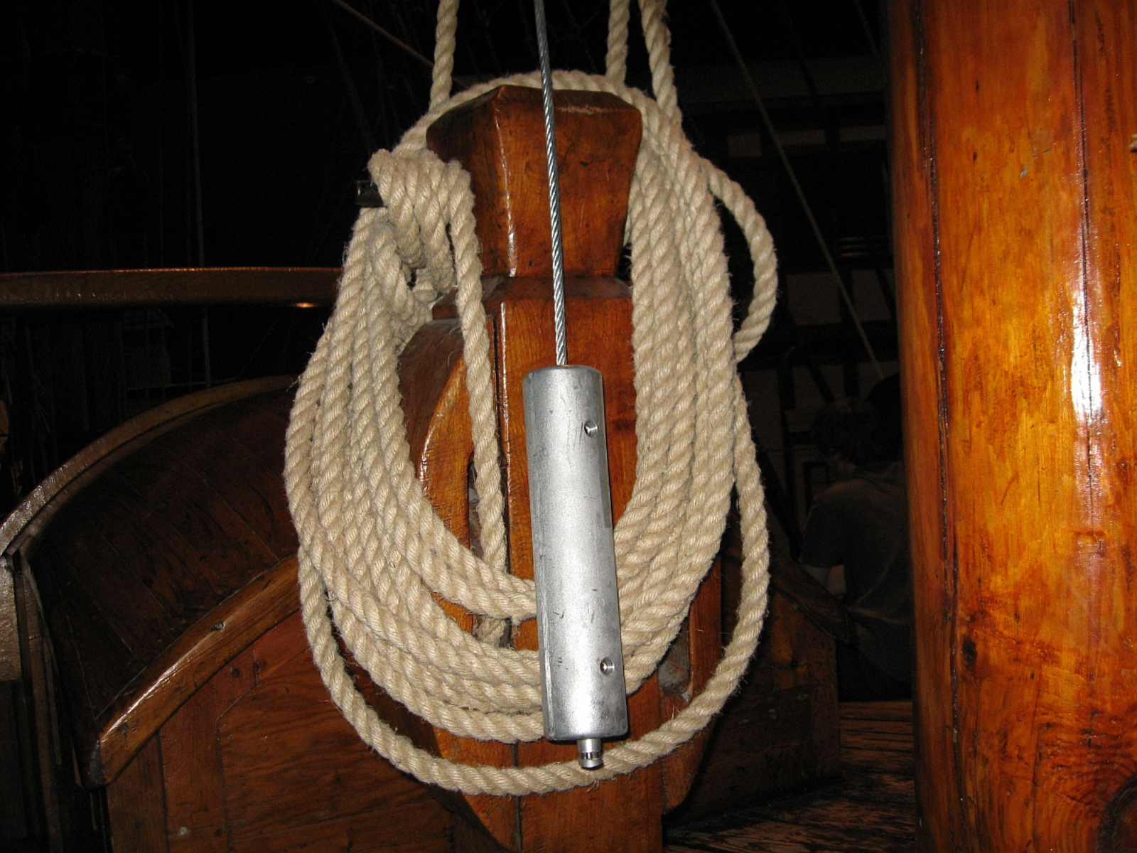
Look up, way up… at the dust on the Nonsuch’s rigging and spars. Museum staff have not been able to climb the ship rigging since Manitoba Workplace Safety and Health regulations were amended several years ago. In order to address the stricter requirements, steel safety cables were installed in the Nonsuch Gallery two years ago. However, they turned out to be less usable than we hoped.

As part of a Conservation Assessment, the Nonsuch was examined by a team including conservation consultants and a ship rigger. One of the recommendations from the assessment was to switch to rope safety lines, for reasons relating to ease of use during climbing. Another recommendation was to add more safety lines, at different spots on the gallery ceiling.

A coil of cream rope on a wooden notch. In the front there's a steel safety line.
A view of one of the original steel safety lines.
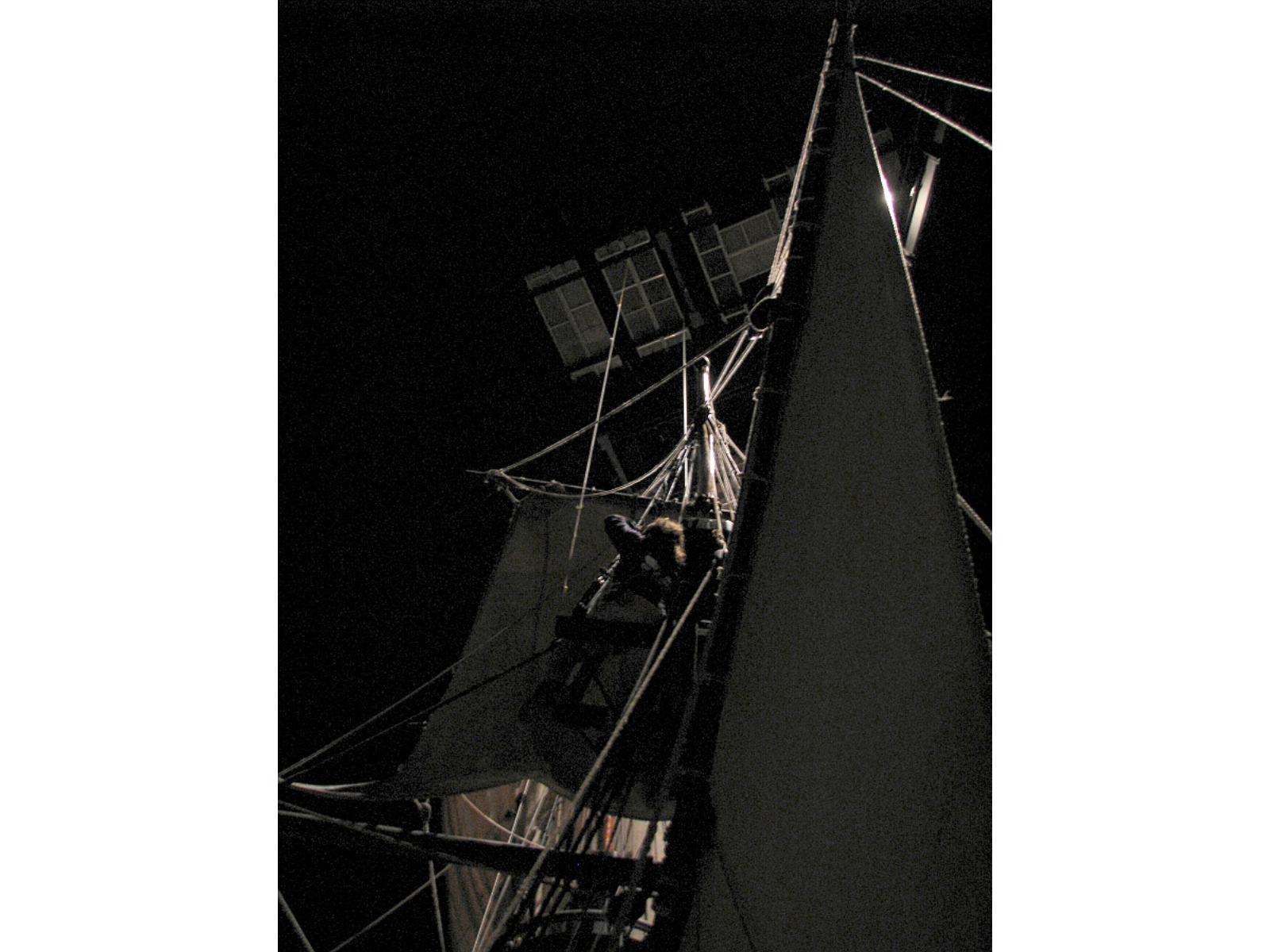
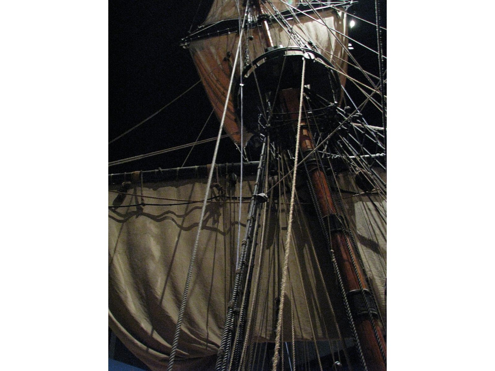
View up into the ratlines and sails of a wooden sailing vessel in an indoor gallery. In the darkness above, the base of a temporary platform can be seen

Dropping a plumbline from temporary platform at ceiling.

The new locations were confirmed, the anchors installed and new synthetic rope safety lines attached. The last task remaining is to do a Risk Assessment and Job Hazard Analysis. After that, we’ll be able to once again climb in the rigging, to do some much-needed cleaning of the lines and sails. Because it’s been several years, we need to plan how best to attack the dust.

View up into the ratlines and sails of a wooden sailing vessel in an indoor gallery.

From a distance, the safety lines blend in fairly well.

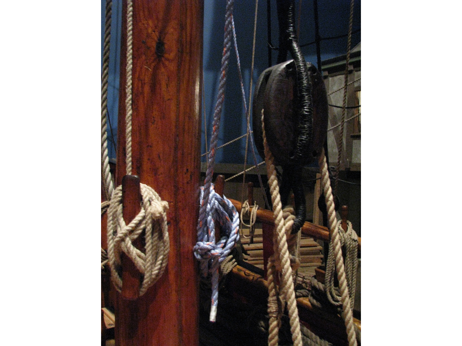
Several coils of rope around notches on a ship's deck. One of the rope's is a more blue-white colour than the others

Close up, they do stand out. I think we’ll move this one somewhere less visible.

The Nonsuch is an unusual thing: a full-sized ship replica, with working rigging, in an indoor setting. There are few other museum ships like it. Although it is an artifact, it is important to keep the rigging at least in working order. Similar to some artifacts in science museums, keeping moving parts static will contribute to their deterioration – ropes will get stiff and develop kinks, making their movement harder and harder, and affecting other parts such as the blocks the lines run through. Keeping the Nonsuch “shipshape” requires regular cleaning and maintenance; soon, one part of that work will be resumed.

True Colours Shining Through – the building of an exhibit

Exhibits begin as the germ of an idea and often take on a life of their own. This was no different for the new Colours in Nature exhibit, now open in the TD Canada Trust Discovery Room. This space was set aside to showcase the hidden treasures of our collections that we ordinarily don’t have an opportunity to display; less that 5% of our collections are exhibited at any one time and there are many spectacular specimens that are available for research, but not generally accessible to visitors.

We (the Natural Sciences section – Botany, Geology & Paleontology, Zoology) had been thinking about ways to combine our collections for exhibit under one theme, but were looking for an additional and different angle to go along with a scientific perspective. What most makes an impact on a behind-the-scenes collection visitor? The numbers of specimens impresses, of course (we have over 150,000 natural history specimens), but it is also often the diversity of colour and beauty of what we have on the shelves that strikes a chord.

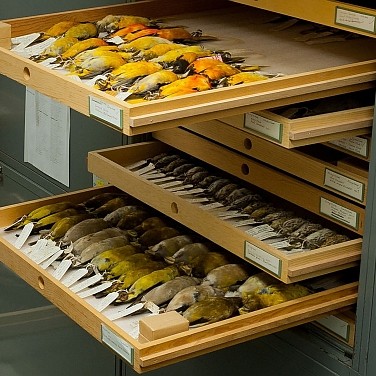
Open storage drawers containing rows of yellow bird specimens.

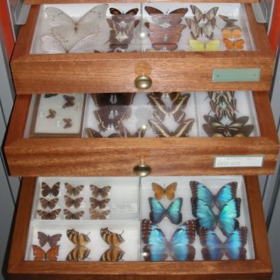
Open storage drawers containing carefully preserved butterfly specimens of various colours, including bright blue.

Trays of blackbirds, orioles and grosbeaks (left) and various tropical butterflies (right), some of the thousands of specimens that were examined as possible exhibition pieces.

So why not have an exhibit on colour? Initially we aimed to create a series of exhibits, each featuring a specific colour, but despite our substantial collections it just wasn’t possible to gather enough specimens of any one colour to make a significant exhibit. We decided on a single exhibit of seven cases, one each for red, orange, yellow, green, blue, purple, and one for multi-coloured specimens. Although the main impact (we hope) would be visual appreciation of the collections, panel text would tie the individual cases together under themes of where colour comes from and how different animals perceive colour. These themes are constrained by text length limits – it is surprisingly tricky to write entertaining, concise, yet meaningful text (haiku, good haiku anyhow, is tougher to write than a full-length novel, in my opinion).

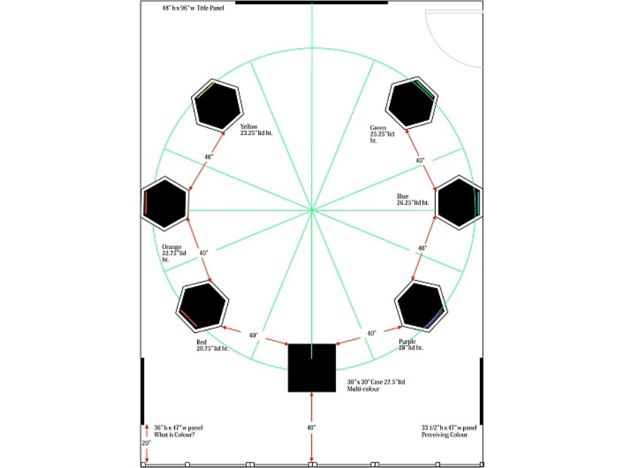
Our concept was shared among other exhibit team members that include an exhibit manager (who keeps us on track and within budget), a designer, conservation and collections personnel, technical support, and programming staff. Exhibit case sizes, arrangement and lighting were determined first, all physical limitations that impact choice of specimens.

Floorplan for the colours exhibit showing seven podiums, one for each of the colours featured.

The floor plan for the exhibit with the square multicolour case front and centre with the hexagonal cases arranged as a colour wheel of red, orange, yellow, green, blue and purple. Black bars on the walls are text panel positions.

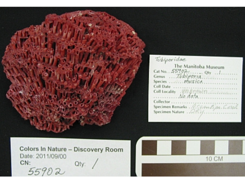
A red coral placed against a black background next to documentation with the object details.

Standardized photos and written records are maintained to track the condition of each specimen over the exhibition period.

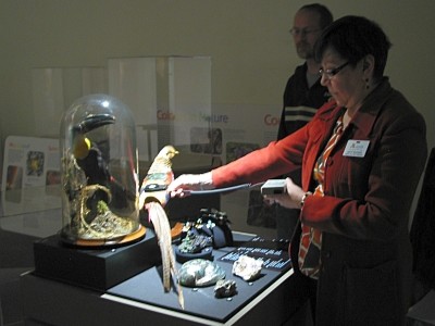
The curators have the onerous, though perhaps enviable, task of going through the collection and choosing appropriate specimens. Along with space limitations, specimens might be unsuitable for display because they are too delicate or are compromised in some way. Each choice is given a final ‘health-check’ by the collections manager through a condition report that includes a photograph; this makes certain that the specimen can withstand the rigors and risks of display and allows us to keep track of any possible deterioration during the course of the exhibition. Lighting and environmental conditions of exhibition galleries can potentially damage specimens.

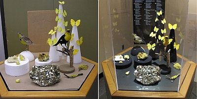
Two photos side-by-side of the yellow exhibit. On the left is a mockup of how the exhibit will look. On the right is the final layout in the exhibit.

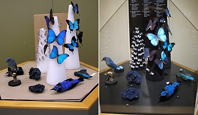
Along with the designer, the curators determine the arrangement for a particular case so that the specimens show their ‘best side’ and the case as a whole is visually interesting. Because Colours is, in a sense, showing the jewels of the collections both figuratively and literally, we were seeking a jewelry display case atmosphere where the colours and vibrancy of the specimens would jump from the cases. We also wanted the colours of the various animals, minerals and plants to form an organic whole where the colours and forms would complement one another.

Two photos side-by-side of the blue exhibit. On the left is a mockup of how the exhibit will look. On the right is the final layout in the exhibit.

Conservation staff finishes the mounts and determines acceptable light levels that will not damage the specimens – all are susceptible to fading. Then curators and collection manager install the mounts,  specimens and labels, and final lighting adjustments are made by technical staff. Designed and proofed panels are wall-mounted. The end result is many hours of individual effort and teamwork.

Only the visitor can judge how successful our efforts have been, but the opportunity is there to see some splendidly brilliant specimens that provide a glimpse into the diversity of our research collections. The final tally for Colours in Nature approaches 300 specimens, and all continents but Antarctica have representatives. With the rare exception, almost all of these specimens are on exhibit for the first time.

Two conservation staff people examining the layout of a portion of the exhibit.

After initial installation, light levels are tested with and without the plexiglass covers to ensure specimens will not sustain damage over the duration of the exhibit.

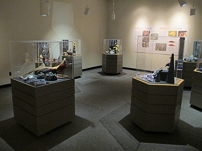
A wide view of several of the set-up podiums for the Colours in Nature exhibit.

A partial view of the final installation showing several of the cases and a wall panel. Each case is lit individually to display the specimens to best advantage.

The exhibit, I think, affirms the words of a Cindi Lauper song, “True colours are beautiful, like a rainbow” – or perhaps, as an educational institution, we would be better to quote Oscar Wilde:

“Mere colour, unspoiled by meaning, and unallied with definite form, can speak to the soul in a thousand different ways.”

Come by this winter and have the jewels of the natural history collections speak to your soul. Although we’d like to think you can leave with a little meaning mixed in there, too.

Dr. Randy Mooi

Dr. Randy Mooi

Curator of Zoology

Dr. Mooi received his Ph.D. in zoology from the University of Toronto working on the evolutionary history of coral reef fishes. Following a postdoctoral fellowship in the Division of Fishes of the Smithsonian Institution…
Meet Dr. Randy Mooi

Cover Shot

By Dr. Graham Young, past Curator of Palaeontology & Geology


Around the Museum this morning, people are excited that Justin Bieber and Selena Gomez visited over the weekend, enjoying a private dinner aboard the Nonsuch. I am pleased that they liked the Museum, and that they were particularly interested in Ancient Seas. But there is another piece of external attention that I am just as pleased about, even if it is unlikely to ever attract a story on Entertainment Tonight. In fact, I would have to say that I am “chuffed.”

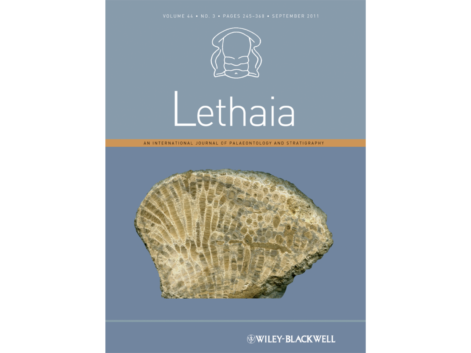
A couple of weeks back, my colleague Bob Elias and I were contacted by the editors of the paleontological journal Lethaia, who were wondering if we might have photos suitable for their cover. It was time for a change from the ammonoid that had graced the cover for several years, and since there were going to be papers about fossil corals and reefs in a coming issue, they were looking for a suitable image of a Paleozoic coral.

Cover mock-up of a book in blue and grey shades with the title, "Lethaia", on the front. A fossil specimen image is positioned front and centre.

So Bob and I scouted around to see what we had. I had some very good photos of Manitoba specimens, but they were all colour slides shot in pre-digital times and I knew from experience that it is hard to get a really first-rate image from a scanned slide. So I delved into the collection, pulling out some of those same specimens and placing them onto the flatbed scanner (read more in blog “On My Scanner”, here). With the scanner the pixel count is virtually infinite, and after a bit of editing I was able to get images that seemed to work. Bob and I selected a variety of photos from what we had, and sent them off to the editors.

Last week we received a message with this cover mock-up:

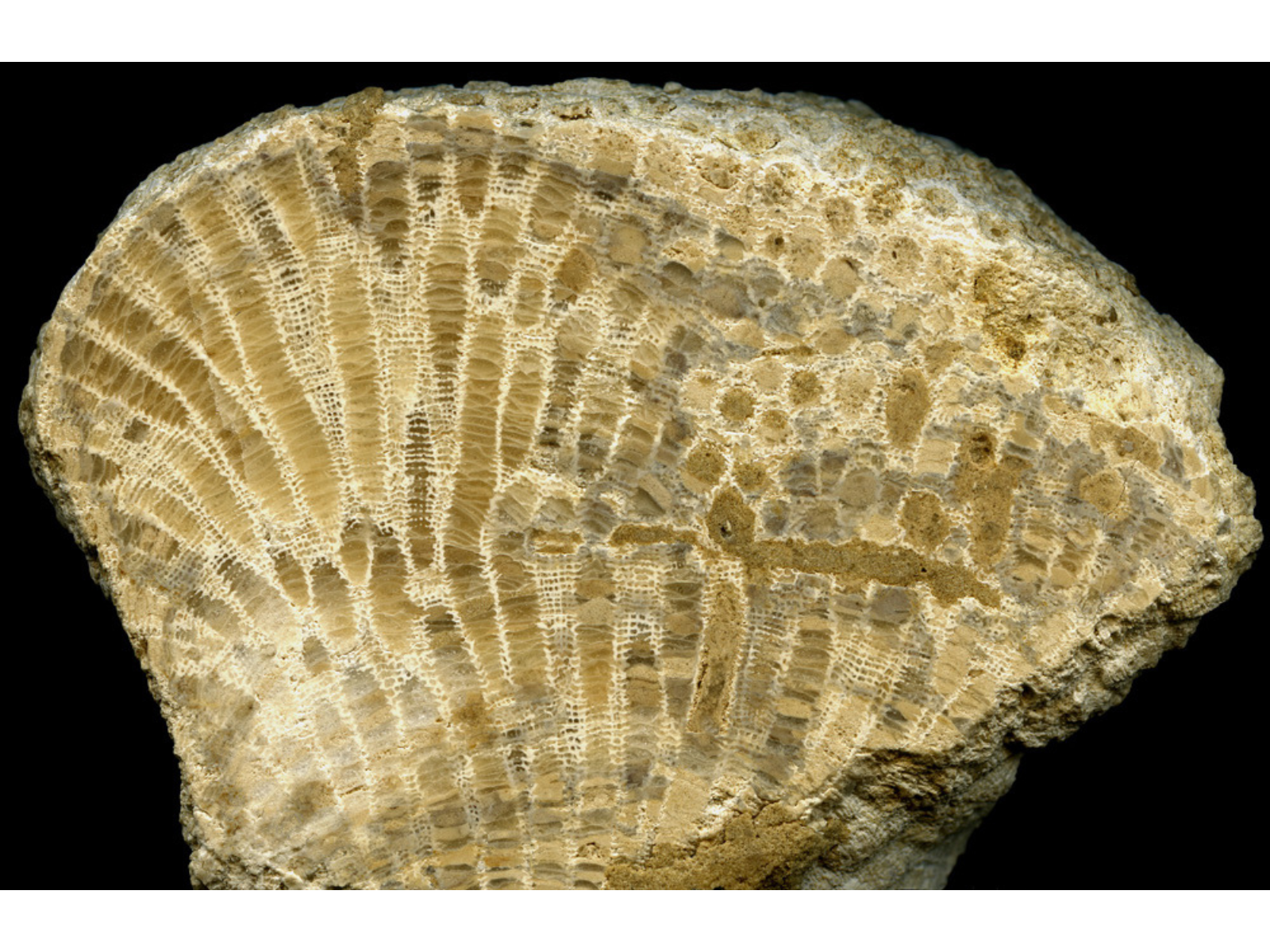
Close-up on a half oval-shaped fossil specimen with a clean cut along the front side showing numerous fossils in the specimen.

There, snuggled into the standard Lethaia cover, is one of the Ordovician tabulate corals from Garson, Manitoba. This is a coral generally identified as Calapoecia sp. cf. C. anticostiensis Billings; the image is of a colony that had been vertically cut and fine polished. The rock unit in which it occurs is the Upper Ordovician (Katian) Selkirk Member of the Red River Formation. This unit, more commonly known as Tyndall Stone, is quarried at Garson and used as a beautiful building stone all over Canada. The coral specimen actually came from rubble heaps at the stone quarry; what could possibly be more representative of this region?


Image: This vertically cut and polished colony of Calapoecia records growth of the coral animals on the ancient tropical seafloor. The horizontal band of sediment to the right of the middle represents an interval in which the animals in part of the colony had died off. This was followed by regeneration as new polyps grew to colonize the “dead” surface (The Manitoba Museum, I-3413).

Maybe this won’t attract hundreds new visitors to the Museum, but it is still nice to have since it will make our existence known to people in very distant places. It is good to see our collections out there; they are so often useful in ways that we have not even thought of!

Colours of Lichens

On October 28 a new natural history exhibit on the Colours in Nature will open in the Museum’s Discovery Room. Organisms and minerals representing all the colours of the rainbow will be on display. Unfortunately no flowering plant specimens will be displayed (only photographs) because, unfortunately, most of the plants in our collection represent only one colour: brown. If the exhibit was on the colour brown in nature, boy would I have a lot stuff to show!

The plants weren’t always brown of course. Before they were collected the leaves were bright green and the flowers hot pink, yellow, orange, and purple. Sadly the drying process almost always results in the loss of at least some colour. Age doesn’t help matters either, the older a plant is, the browner it gets.

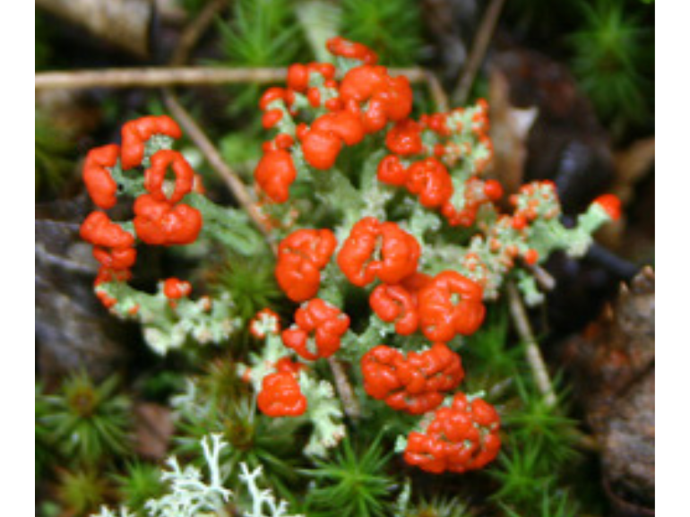
Close-up on a cluster of small mushrooms with bulbous, waxy red caps.

British Soldier lichens have distinct red caps.

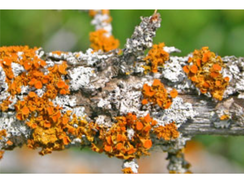
Textured orange lichen growing alongside smooth silvery lichen on a branch.

Gold-eye lichens are becoming rare because they are sensitive to air pollution.

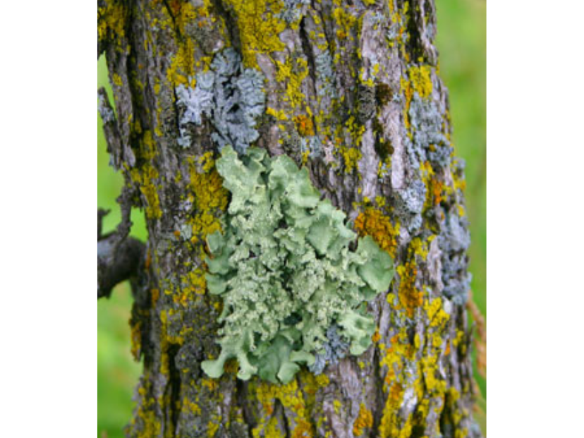
Patches of green, yellow, and silvery lichen growing on a tree trunk.

The specimens from the Botany collection that ARE on display are fungi, lichens in particular. Lichens are a fascinating group of organisms. They consist of tiny algae or cyanobacteria (the photobiont) living inside the tissue of a fungus (the mycobiont). The relationship is, for the most part, mutually beneficial with the algae photosynthesizing and providing carbohydrates to the fungus and the fungus protecting the algae. This symbiosis enables both organisms to live in harsh environments where they probably couldn’t live independently (like on rock). Some lichens living in really harsh environments (like the Antarctic) are cryptoendoliths, meaning that they live inside the rock, penetrating the tiny spaces in between granite and marble crystals. In fact, some scientists think that if we find life on Mars it may be inside the rocks!


Image: Green, yellow and silvery lichens are common on oak trees.

In some lichens the colour of the green algae inside the fungus shows through, especially when wet. If the lichen dries out, it may look grey or brown. Other lichens produce brightly coloured pigments like the brilliant yellow pulvinic acid or the reddish anthraquinones. Why lichens produce these chemicals is still a bit of a mystery but one of the best hypotheses is that they act as algal sunscreen, protecting the sensitive algae from harmful UV radiation in sunny habitats. For this reason, the brightest lichens in Manitoba can be found on the tundra in the far north as well as on rocks in the grasslands.

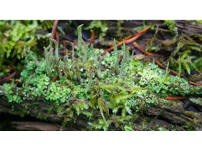
Vibrant green, low-growing lichen with algae growing inside.

The green colour of the algae inside this Cladonia lichen shows through when wet.

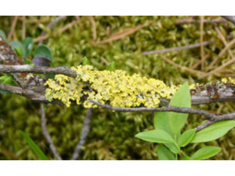
Yellow lichen growing on branches near the ground.

Powdered sunshine lichens were traditionally used as a source of yellow dye.

So if I’ve piqued your curiosity, come on down to the Museum to see our collection of brightly coloured lichens (and minerals and brightly coloured birds and butterflies too).

Dr. Diana Bizecki Robson

Dr. Diana Bizecki Robson

Curator of Botany

Dr. Bizecki Robson obtained a Master’s Degree in Plant Ecology at the University of Saskatchewan studying rare plants of the mixed grass prairies. After working as an environmental consultant and sessional lecturer…
Meet Dr. Bizecki Robson