

Posted on: Wednesday October 30, 2013
Hallowe’en is upon us and all the traditional ghosts, goblins, witches, and bats are making their annual appearance. The Museum just hosted a very successful members’ night that included trick-or-treating for kids. But a recent (unfortunate) offer of a real bat to our Zoology collections has me thinking that we need to re-evaluate the inclusion of bats as a Hallowe’en symbol – they just don’t belong at this time of year!
Manitoba has six species of bats. Half of these are “cave bats” as shown in the image below to the left. Over the winter, most of the individuals of these species likely stay inside the province or in nearby provinces or states by hibernating in caves. Big brown bats occasionally find shelter in buildings. The caves are cool, but do not freeze and offer stable temperatures and humidity that are good for hibernation.
The other three species are the “tree bats” that are illustrated in the image below to the right; the red bat is a particularly attractive species (although for many, “attractive bat” is likely considered an oxymoron!). The “tree bats” escape our winters by migrating out of the province in late summer and fall, likely to the southern United States, and return each spring.
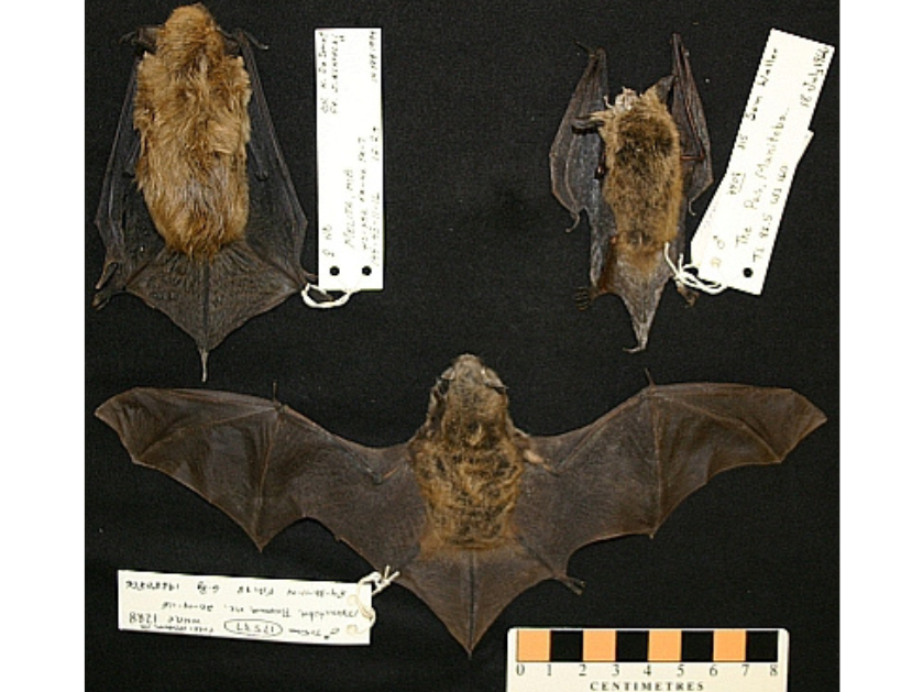
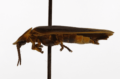
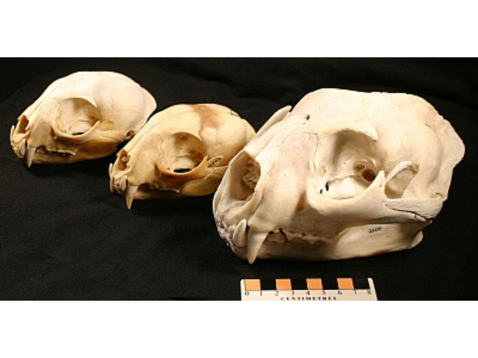
The three “cave bats” of Manitoba: big brown bat (Eptesicus fuscus, MM 17638, 12.2 g, Melita), upper left; northern long-eared bat (Myotis septentrionalis, MM 4408, 7g, The Pas), upper right; little brown bat (Myotis lucifugus, MM 17537, 6.8 g, Pinawa), bottom. These hibernate in caves over winter.
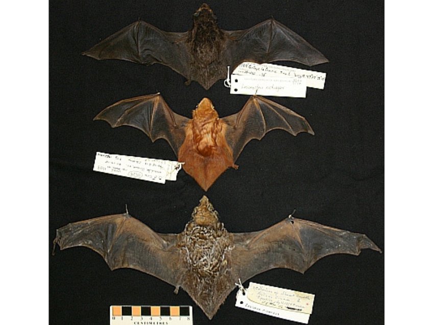
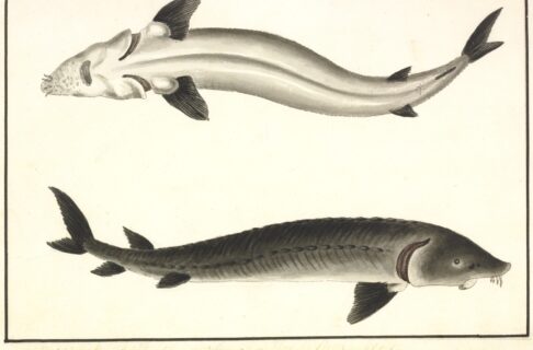
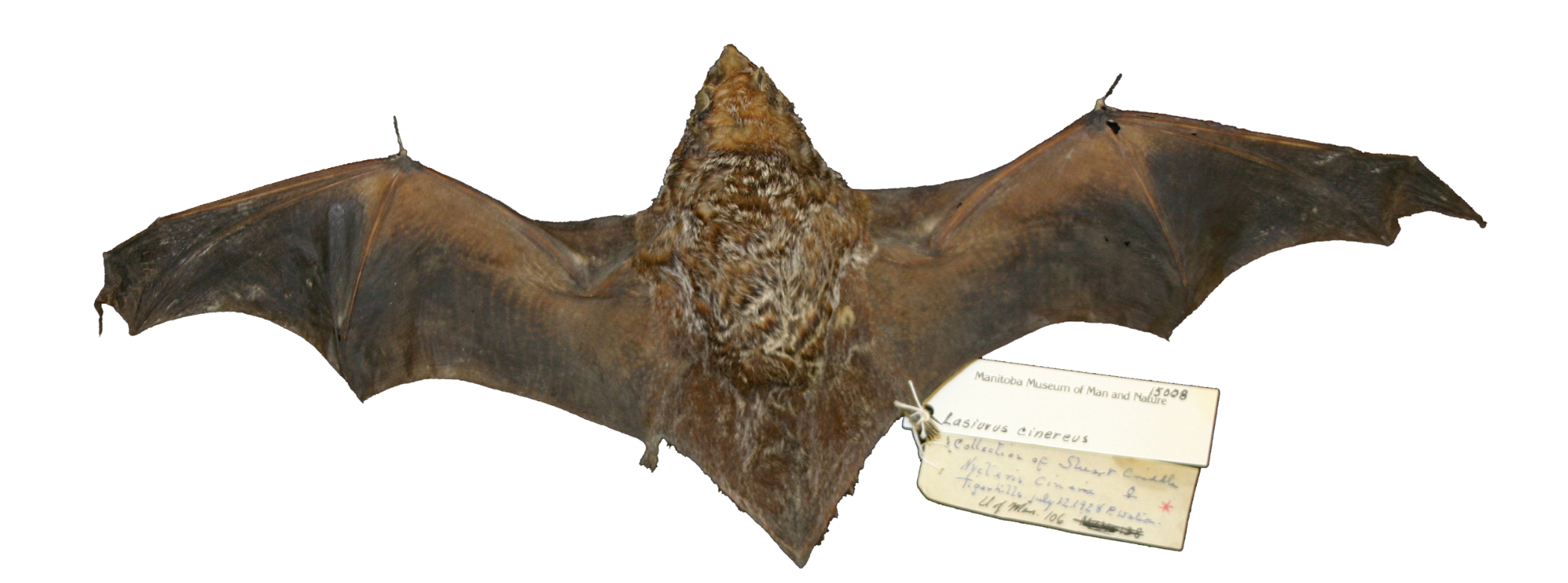
The three species of “tree bats” in Manitoba (from top to bottom): silver-haired bat (Lasionycteris noctivagans, MM 9972, 11.7 g, Portage La Prairie); eastern red bat (Lasiurus borealis, MM 17529, 9.5 g, Pinawa); hoary bat (Lasiurus cinereus, MM 15008, 27.8 g, Tiger Hills). These species migrate south to avoid our winters.
The other three species are the “tree bats” that are illustrated in the image above; the red bat is a particularly attractive species (although for many, “attractive bat” is likely considered an oxymoron!). The “tree bats” escape our winters by migrating out of the province in late summer and fall, likely to the southern United States, and return each spring.
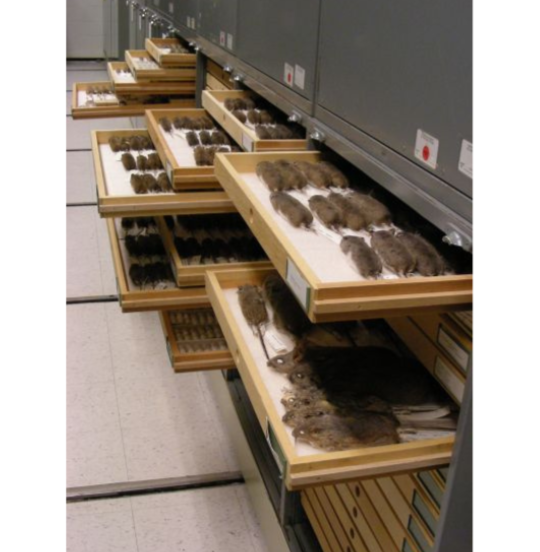
But just as with us when we wait too long to put on the snow tires, bats sometimes get caught by cold weather. That is what happened to an unfortunate silver-haired bat just last week (October 23) that was found lying dead on a Winnipeg sidewalk. Despite attempts at warming the little guy, it did not revive and was offered to be a part of the research collection here at the Museum. Such donations provide the raw materials to help understand these (and other) little-known animals. The Museum is grateful for these donations, and in some ways, gives the organism a second “life” where it can be studied and be used for exhibits and other educational purposes.
The silver-haired bat is likely the commonest tree bat in Manitoba (and North America), but despite this we know surprisingly little about it. They are long-distance migrants, probably spending the winter in the southern U.S. and returning to Manitoba certainly by May – we have found them clinging to the Museum on occasion as they are on their way to northern forests. These are spectacular trips for animals that weight the equivalent of $2 in loonies! [That’s about 12 g.] In summer, the species is found in north temperate zone conifer and mixed conifer/hardwood forest feeding mostly on soft-bodied insects, particularly moths, but also midges and mosquitoes. Females form small, communal maternity roosts in tree hollows or under bark. In August and September they migrate back south to avoid our cold weather and lack of insects.
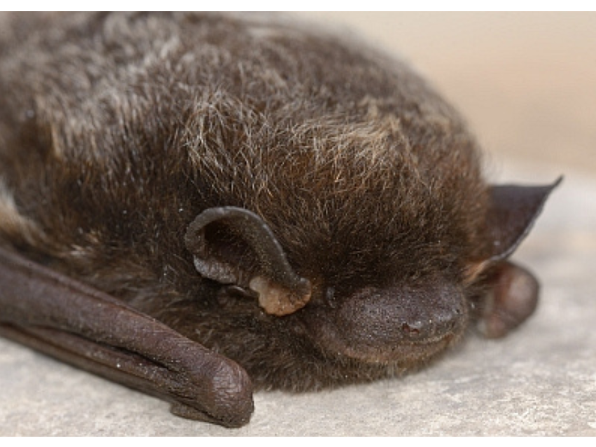
The face of a silver-haired bat (Lasionycteris noctivagans). This little fellow (it was a male) waited too long to leave the province and was found dead on a Winnipeg sidewalk. He will be donated to become part of the research collection, available to provide a little more knowledge about these poorly-known mammals. Photo provided by D. Dodgson, and published with permission.
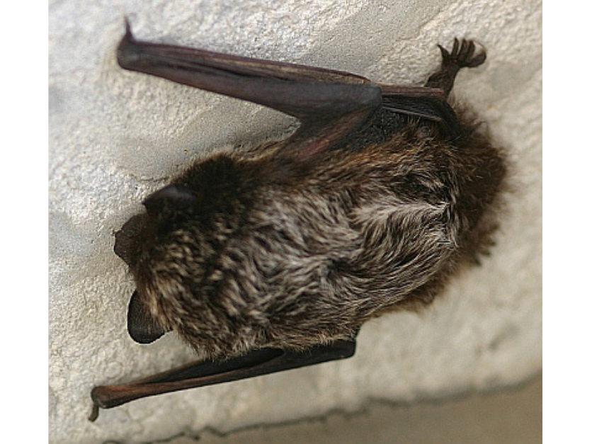
A silver-haired bat (Lasionycteris noctivagans) clinging on to the Manitoba Museum during migration in May, 2008. This bat had disappeared by the following morning, migrating further north.
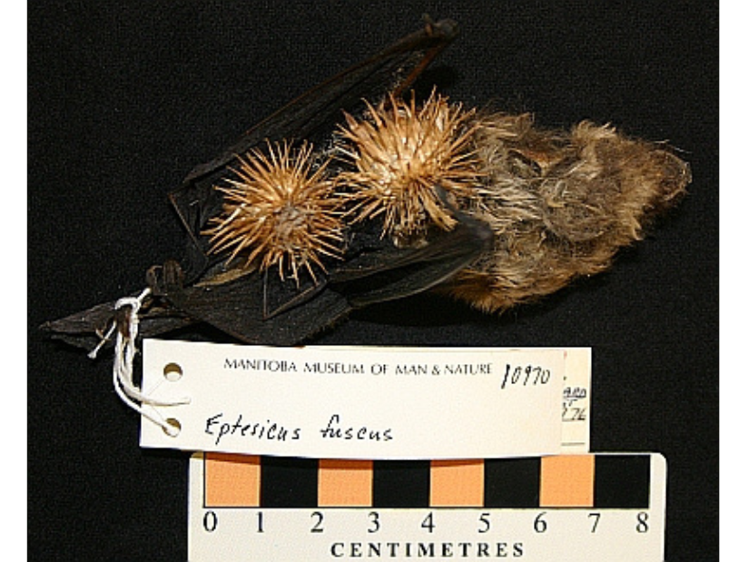
Cold weather is not the only hazard to bats as they migrate to find caves or move south. This big brown bat (MM 10970, Winnipeg) was caught by the hooked fruit of a burdock plant and could not escape.
Knowing that Hallowe’en frequently seems like the coldest day of fall – it always seemed so to me as a parent taking kids around the neighbourhood! – all of our bats should either have found safe hibernation sites or have moved south by now. Most silver-haired bats should have left at least a month ago (end of September), long before our local ghosts, goblins, and witches start wondering our streets. And given that October weather is so hard on these little mammals, with the recent offering to the Museum collection as clear evidence, Hallowe’en and bats just don’t mix.
In Manitoba, Hallowe’en is a far scarier time for bats than it is for any of our trick-or-treaters.